
A Standard Operating Procedure SOP for Identification of Unknown Microorganisms is crucial in a microbiology laboratory to ensure consistency, accuracy, and safety in the process. Keep in mind that specific procedures may vary based on the laboratory’s equipment, resources, and protocols.
PURPOSE
To describe the procedure for the identification of bacteria as Gram Negative and Gram Positive up to specie level
SCOPE
This procedure is applicable to elucidate the morphology arrangement of bacterial cells.
RESPONSIBILITIES & AUTHORITIES
- Manager Quality Control is responsible to ensure that procedure & formats are followed entirely as approved.
- Microbiologist is responsible to perform the test.
REFERENCES & REQUIREMENTS
USP
DEFINITIONS & ABBREVIATIONS
Gram Staining:
A staining method used to classify different species of bacteria.
Principle SOP for Identification of Unknown Microorganisms
A Danish physician, Hans Christian Gram developed this staining procedure in 1883. It is differential staining method by which bacteria can be classified into two broad groups. The bacteria which retain the color of primary dye even after treatment with alcohol, are termed as Gram Positive and those which are decolorized by alcohol and take the color of secondary dye are known as Gram Negative. Gram reaction depends upon the chemical nature of the cell wall of the bacteria. In Gram positive, the concentration of lipids is much lower (1 – 4%) than the Gram negative (11 – 22%). The lipids in Gram negative are dissolved in alcohol, thus making their cell wall more permeable. This leads to an outward flow of primary dye from the cell rendering it colorless. Such cells take the color of secondary dye. In the contrary, the lipid content of Gram positive bacteria does not affect the permeability of the cell wall and thus the primary dye does not leave the cell. Gram staining is of great significance in the diagnosis of various diseases and in the identification of bacteria.
Gram-positive bacteria:
Gram-positive bacteria are those that are stained blue violet by staining. They have a thick layer made up of polymers of protein-sugar molecules called peptidoglycan.
Gram-negative bacteria:
Gram Negative bacteria are those which cannot retain the crystal violet stain, instead taking up the counter stain (safranin or fuchsine) and appearing red. They have an additional outer membrane whose properties resist acetone/alcohol-dehydration and result in loss of the crystal violet stain. A counter stain, made up of fuchsin, stains these bacteria red or pink.
ABBREVIATIONS
SOP – Standard Operating Procedure
SCDA – Soybean Casein Digest Agar
LAF – Laminar Air Flow
XLDA – Xylose Lysine Deoxycholate Agar
BSA – Bismuth Sulphite Agar
VJA – Vogel Johnson Agar
BPA – Baired Parker Agar
CA – Cetrimide Agar
VRBG – Violet Red Bile Broth
SS Agar – Salmonella Sheigella Agar
PROCEDURES SOP for Identification of Unknown Microorganisms
Materials & Equipment’s
General Requirements:
- Cavity slides.
- Cover slips.
- Bacterial cultures
- Alcohol (70% IPA)
- Tooth picks.
- Bunsen Burner
- Immersion oil
- Microscope
Preparation of Gram Staining Reagents:
Preparation of Crystal-violet solution:
- Crystal Violet 2.0 g
- Ammonium Oxalate 0.90 g
- Ethanol 9.5 ml
- Distilled water (q.s) 100ml
Preparation of Lugol’s solution (iodine potassium- iodide solution):
- Iodine 1.0 g
- Potassium iodide 2.0 g
- Distilled water (q.s) 100mL
Preparation of Decolorizing Agent (Acetone Alcohol):
- Acetone 50 mL
- Alcohol (96 %) 7.5 mL
- Distilled water 2.5 mL
Preparation of Safranin Reagent:
Safranin 2.5 g
Alcohol (96 %) (q.s) 100 m
Preparing the specimen:
One drop of the bacterial suspension is spread in the middle of a cleaned and degreased microscope slide. The SOP for Identification of Unknown Microorganisms liquid is dried cautiously in the heat a couple of centimeters above a low gas flame, where after the preparation is fixed by moving it quickly a couple of times through the upper part of the flame, the side with the preparation being turned upwards.
Staining with crystal violet:
Crystal violet solution is poured the preparation (reaction time: One minute).
Treatment with iodine:
The crystal violet solution is poured off, and lugol’s solution is poured on, so that the latter mixes quickly with the remaining crystal violet solution; after decanting fresh lugol’s solution is poured on (in all one-minute treatment).
Rinsing with Decolorizing Agent:
The microscope slide is kept slanting, and decolorizing agent is poured on, in such a way that the alcohol weeps the lugol’s solution away, without the two liquids’ having an opportunity to mix. After pouring in fresh decolorizing agent, the preparation is shaken for about 30 seconds, in order that the decolorizing agent can act uniformly.
Rinsing with water:
The slide is again kept slanting and is rinsed with water in such a way that the water sweeps away the alcohol, without any mixing of the two liquids.
Staining with safranin reagent:
After having let the water drain off thoroughly, a staining with safranin reagent is carried out for 2 minutes.
Rinsing and drying:
The preparation is rinsed with water and dried cautiously by pressing between two layers of dried filter paper.
Microscoping:
The stained preparation is examined in the microscope using immersion oil.
Results:
Gram-positive bacteria appear bluish black, while gram-negative bacteria are red. (If Doubtful intermediate states are found, the staining has not been carried out correctly, in which case it must be repeated.)
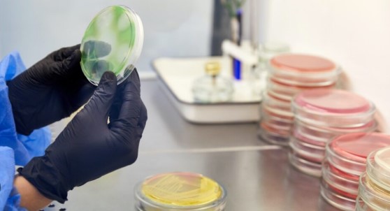
Verify the colony characteristics of microbial culture in respective media.
If colony characteristics do not match the point. Record the morphology only.
Lactophenol Cotton Blue Staining for Fungi
- Aseptically open microbial culture tube under LAF.
- Take a clean grease free slide.
- Take the mycelium (for filamentous fungi) of the fungi with the help of needle and forceps and place it on the slide. Carefully tease the mycelium and add few drops of Lactophenol cotton blue cover the whole mycelium, cover with a clean grease free coverslip and observe under the microscope.
- Compare the morphology of the culture observed in the microscope with the morphology mentioned in the point.
- Verify the colony characteristics of microbial culture in respective media as per point.
- If colony characteristics do not match the point. Record the morphology only.
- SOP for Identification of Unknown Microorganisms
Morphology of Microbial Culture
- E. coli: Coccobacilli Gram’s Negative –Indole production- positive (Colonies with Green Metallic Sheen on EMB agar and Pink to dark pink dry donut shaped colonies on Macconkey Agar)
- Salmonella sps: Gram’s Negative rods –Urease negative (Black colonies on XLDA, BSA)
- Staphylococcus aureus: Gram’s positive cocci in bunches-Coagulase positive(Black colonies with yellow zone on VJA, Black colonies with clearing zone on BPA)
- Pseudomonas aeruginosa: Gram’s negative short rods –motile, Oxidase positive (Fluorescent green color on CA)
- Bacillus subtilis: Gram’s negative Bacilli spore-forming, non-capsulated, motile. (Non-mucoid, Irregular colonies on SCDA)
- Aspergillus niger: Filamentous fungi on microscopically show branching and septate hyphae. (Black spore-forming fungi)
- Candida albicans: Yeast Like/ Dimorphic Fungi oval/spherical budding yeast produces septate pseudohyphae & true hyphae (Whitish cream colored mucoid large colonies)
- Shigella. flexeneri: Gram’s Negative rods (Off-white to colorless colonies on SS Agar)
- Lactobacillus plantaurm: Noncapsulated Gram’s positive rods which tend to occur singly or in chains (Shows heavier cells in Pentothenate inoculum Broth).